Helpful Articles

Helpful Articles

Helpful Articles

Helpful Articles


Our eyes are extremely delicate, yet they can be subjected to harsh conditions and other environmental factors that affect their health. One of the problems that can affect our eyes is an accumulation of dirt, debris and bacteria on the eyelids. This can cause a range of issues, including stopping tear film from reaching the eyes and being properly dispersed over their surface – which is necessary to keep them healthy and comfortable. Fortunately, a new solution called BlephexÔ can help.
What is BlephexÔ?
BlephexÔ is a handheld electro-mechanical device that is applied to the margins of the eyelids with the purpose of cleaning them and improving the effectiveness with which tear film flows onto the surface of the eyes.
BlephexÔ has a disposable, surgical-grade sponge tip which rapidly oscillates to create a cleaning action. Before the sponge tip is placed onto the eyes, it is soaked in a gentle exfoliating solution. This solution provides soft abrasion to help remove dead skin cells and debris that could be irritating the eyes and interrupting tear film progression. The BlephexÔ device is manually applied to the eyes and moved gently across the eyelids, with the entire, painless process taking approximately 6 to 8 minutes per eye. A different sponge is used on each eye, ensuring that no bacteria is passed between them. After the procedure, patients are given instructions on how to maintain the cleanliness of their eyelids with daily/nightly eyelid hygiene at home.
Most patients experience a significant improvement in tear film production and dispersal, and a reduction in unpleasant symptoms that they may have been experiencing within 48 hours of their treatment. While a single treatment is normally enough to produce excellent results, many patients are advised to have BlephexÔ every 4-6 months.
What conditions can BlephexÔ help with?
BlephexÔ can be used to clean the eyelids at any time, and people who suffer from dry eyes or eye allergies may find it is particularly beneficial for helping to reduce the symptoms that they experience. It can also be combined with Lipiflow – another technological solution – to help counteract the effects of dry eyes.
Unsurprisingly, BlephexÔ is particularly recommended as a treatment for an eye condition called blepharitis. Blepharitis is characterized by the inflammation of the eyelids, which causes them to become red, swollen and itchy. Although the condition is not usually serious, it can lead to further problems if it isn’t treated.
Symptoms of blepharitis include:
Sore eyes
Itchy eyes
A gritty, irritated feeling affecting the eyes
Redness
Flakes or crustiness around the roots of the eyelashes
Eyelids that stick together when you wake up in the morning
If you are suffering from the symptoms of blepharitis, dry eyes or eye allergies and feel that you would benefit from BlephexÔ treatment, please contact our team to schedule a consultation appointment.

Ask Our Eye Doctor in Richmond, Virginia, how To Prevent Vision Loss
Vision loss is more common than you may think! In fact, it's among the most prevalent disabilities in adults and children. Knowing what puts you at risk of developing vision loss is important and can help you to be proactive about caring for your eyes.
Below, we’ll explore the most common causes of vision loss and the risk factors associated with each.
Spreading awareness and education about visual health is just one way that our eye doctors near you can help. To schedule your Comprehensive eye exam, call us today (804) 285-7638.
Glaucoma
Glaucoma is a group of eye diseases caused by a buildup of pressure within the eye. Too much inner-eye pressure can damage the optic nerve and lead to vision loss.
Since symptoms don't usually manifest in the early stages of glaucoma, getting regular eye exams is all the more crucial. Advanced or rapidly progressing glaucoma can show a variety of symptoms, such as blurred vision, headache, severe eye pain and redness, seeing halos around lights, and nausea.
Risk factors for developing glaucoma include:
- Being 60 years or older
- Family history of glaucoma
- African, Asian, or Hispanic descent
- High myopia (nearsightedness) or hyperopia (farsightedness)
- Previous eye injury or certain eye surgeries
- Certain medications, like corticosteroids
- Thin corneas
- Certain medical conditions, like diabetes, hypertension, heart disease, and sickle-cell anemia
Cataracts
Cataracts occur when the eye's lens becomes cloudy. A healthy lens is clear and allows light to pass through it undisturbed.
Common cataract symptoms include cloudy or blurred vision, difficulty seeing at night, light sensitivity, double vision in the affected eye, and seeing colors as faded or yellowish.
Risk factors for developing cataracts include:
- Aging
- Diabetes
- Hypertension
- Smoking
- Previous eye surgery, injury, or inflammation
- Alcoholism
- Extended use of corticosteroids
Age-Related Macular Degeneration (AMD)
AMD is the leading cause of severe vision loss in adults over the age of 60. It occurs when the macula (the small central portion of the retina, which is responsible for sharp, colorful, central vision) begins to wear down.
Early stages of AMD usually go unnoticed, but later stages of the disease can produce symptoms like blurred vision, dark or blurry areas in your central vision, and problems with color perception.
There’s not yet a cure for AMD, but certain treatments can help prevent vision loss.
Risk factors for developing AMD include:
- Smoking
- Obesity
- Aging
- Long-term sun exposure
- Hypertension
- Heart disease
- Family history of AMD
- Light-colored eyes
- Farsightedness
Diabetic Retinopathy
Diabetic retinopathy (DR) is a complication of Type 1 or 2 diabetes that affects the light-sensitive tissue at the back of the eye called the retina.
Initially, diabetic retinopathy shows no symptoms but can eventually lead to blindness. As it develops, it can cause increased floaters, impaired color vision, dark spots in your visual field, and blurred vision.
Risk factors for developing diabetic retinopathy include:
- Length of time from diabetes diagnosis — the longer you’ve had it, the higher your chances of developing visual complications
- Uncontrolled blood sugar
- Obesity
- High cholesterol or blood pressure
- Pregnancy
- Smoking
- African American, Hispanic, and Native American ethnicities
- Family history of DR
So, what’s the bottom line?
Multiple factors contribute to eye disease and vision loss, and some may even be relevant to you. If you think you may be at risk for vision loss or experience any of the symptoms listed above, speak with your eye doctor in Richmond as soon as possible. We also recommend you have your eyes thoroughly examined every 1-2 years, or as often as your eye doctor recommends. To schedule your comprehensive eye exam, call Patterson Eye Clinic today.
Book an eye exam at an eye clinic near you to learn more about your candidacy for contact lenses and which type is right for you.
Bloodshot Eyes – Should You Be Concerned?

You wake up in the morning ready to start your day, only to discover that your eyes are bloodshot. That might not be surprising if you stayed up late to finish a project, had too many drinks at a party or spent time in a smoke-filled room.
But bloodshot eyes can also signal an underlying eye problem. If your eyes appear red or bloodshot, make an appointment with an eye doctor for a comprehensive eye exam to determine the cause and to receive effective treatment.
Why Do I Have Bloodshot Eyes?
When blood rushes to the front of the eye, the tiny red blood vessels on the white of the eye dilate and become visible. This makes the eyes appear red and irritated.
So why do these blood vessels dilate, causing your eyes to look bloodshot?
Bloodshot eyes tend to be caused by:
- Dry eyes
- Irritants such as smoke, pollen and perfume
- Lack of sleep
- Excessive alcohol consumption
- Spending too much time in front of the computer
Bloodshot eyes due to lifestyle and environmental irritants may disappear on their own, or you can try to relieve them with over-the-counter eye drops or liquid tears. Lifestyle changes, such as getting more sleep, cutting down on alcohol intake and limiting screen time can often be helpful. If allergies are the culprit, oral antihistamines and antihistamine eye drops may relieve symptoms.
At other times, underlying problems requiring prompt medical attention can cause your eye’s blood vessels to dilate. The following are some of these medical conditions:
Conjunctivitis
You’ve probably heard of “pink eye.” It's another name for infectious conjunctivitis – an infection of the conjunctiva, the thin membrane covering the eyelid and the front surface of the eye.
There are two types of infectious conjunctivitis – bacterial and viral.
If your child has conjunctivitis, they’re not alone. About 12% of kids get bacterial conjunctivitis every year. This highly contagious condition affects children and adults. In addition to reddish eyes, the following symptoms are associated with conjunctivitis:
- Bacterial conjunctivitis - irritated eyes, swollen eyelids, eye discharge, crusty eyelids and excessive tearing
- Viral conjunctivitis - cold or flu-like symptoms, runny nose, fever, itchy eyes, excessive tearing
If you or your child are experiencing these symptoms, it’s important to schedule a prompt appointment with an eye doctor, who can diagnose whether the conjunctivitis is viral, bacterial or due to allergies.
Depending on the diagnosis, your eye doctor will prescribe antibiotic eye drops or creams to treat bacterial conjunctivitis. The viral form may run its course after a few days, but cold compresses and non-prescription eye drops may provide relief.
Dry Eye Syndrome
If your eyes are chronically bloodshot you may have dry eye syndrome (DES). Signs of DES include:
- Dry, irritated eyes
- Burning or stinging eyes
- Discharge from the eyes
- Light sensitivity
- A feeling you have something stuck in your eyes
- Blurred vision
- Watery eyes
Dry eye syndrome is most commonly caused by a blockage of the tiny meibomian glands in the eyelids. These glands secrete oil that keeps eye moisture from evaporating too quickly. Without the oil, tears dry fast, leaving your eyes feeling dry, itchy and with a bloodshot appearance.
Too much screen time, aging, certain medications such as antihistamines, and medical conditions such as Sjogren’s syndrome can cause dry eye syndrome.
In addition to any medications or in-office treatments your eye doctor recommends, make sure to get plenty of hydration, take frequent breaks from digital screens and use a humidifier in your home.
Uveitis
In addition to bloodshot eyes, if you also experience blurred vision, see floaters or your eyes feel painful, you may have an eye inflammation called uveitis. The causes of uveitis include:
- Autoimmune or inflammatory condition
- Infection
- Medication side effects
- Cancer (in rare cases)
Unfortunately, uveitis symptoms can often be mistaken for something less serious. That’s the reason it’s important to get an eye exam if your eyes are bloodshot. Left untreated, uveitis can lead to serious conditions such as retinal scarring, cataracts and vision loss.
Depending on the cause and severity, your eye doctor may treat uveitis with prescription eye drops, steroid pills, injections or eye implants.
Eye Injury
It’s vital that all eye injuries receive immediate eye care from an eye doctor.
Even a minor eye injury can cause a big red blotch to form on the white part of the eye (sclera). The cause is a broken blood vessel or a subconjunctival hemorrhage.
Although the appearance of this blood looks severe, and can make the entire white part of the eye appear bright red, a subconjunctival hemorrhage is usually painless and doesn’t cause vision loss. Any time you notice excessive blood on the eye following an eye injury, schedule an appointment with an eye doctor to assess the health of your eye.
Glaucoma
In rare cases, bloodshot eyes may signal the presence of glaucoma – a leading cause of vision loss and blindness.
While some types of glaucoma don't show symptoms in the early phases, bloodshot eyes can indicate the type of glaucoma that requires immediate medical care. This disease causes damage to the optic nerve due to excessive pressure within the eye. When this pressure suddenly rises, the eye’s blood vessels become dilated and visible, making the eye appear red.
If you have bloodshot eyes and/or have the following risk factors for glaucoma, immediately schedule an appointment with your eye doctor.
- Family history of glaucoma
- Aged 60+
- African American, Asian or Hispanic
- Diabetes
- High blood pressure
Bloodshot Eyes Won’t Go Away?
Talk to Us Any time you notice bloodshot eyes or blood on the front of the eye, don’t wait. Schedule your eye exam with Dr. Peter Nardone at Patterson Eye Clinic in Richmond today.
Q&A With Your Local Optometrist
Can I get bloodshot eyes after LASIK surgery?
LASIK surgery is highly effective minimally invasive laser eye surgery that can correct refractive errors, but like all surgical procedures, it can have side effects. Your eyes may be bloodshot or you could see halos from a few days to three weeks after surgery. Additionally, you may experience other dry eye symptoms. Eye drops and liquid tears can alleviate these symptoms, but if you have any concerns about your eyes following LASIK surgery contact your eye surgeon.
What Should I Expect from a Glaucoma Exam?
If you have a family history and/or other risk factors for glaucoma, and if your eyes look bloodshot, consider scheduling a glaucoma exam. Your eye doctor may perform the following tests:
- Tonometry - eye pressure test
- Gonioscopy – to see how fluid is draining out of your eye
- Vision field test – to examine the functioning of the optic nerve
- Dilated pupil exam – to detect any damage to the optic nerve
- Retinal photo or OCT - digital examination of the retina and optic nerve health

Optical Coherence Tomography is a non-invasive imaging test that may be performed as a standard part of your regular, comprehensive exams, or you may be able to request this test as an addition to your usual exam.
Optical Coherence Tomography uses light waves to take cross-section images of your retina, which is the area of light-sensitive cells at the back of your eye that is responsible for receiving light and transmitting it into messages that are sent up to the brain. The technology behind OCT enables your eye doctor to see each of the different layers that make up the retina. By being able to see these and measure them, they can obtain a much clearer picture of the overall health and condition of your eyes.
Why are Optical Coherence Tomography scans important?
When you choose to have an OCT scan at fairly regular intervals, such as during your normal comprehensive eye exams, your eye doctor can compare newer results to previous ones. This helps them to build up a picture of the health of your eyes, and spot any changes which may be concerning, early, before they cause symptoms or have a permanent effect on your vision.
Anyone can have an OCT scan, but they are particularly recommended for patients over the age of 25 who are concerned about the health of their eyes, or who are at risk of or already have diabetes, glaucoma or a family history of eye disease. This is because they can be used to spot the early signs of a range of eye diseases, including glaucoma, diabetic retinopathy, macular degeneration, disorders of the optic nerve and more – even before you realize that you are affected.
What happens during an Optical Coherence Tomography scan?
An OCT scan is a quick, painless experience. To prepare you, your eye doctor may require you to have eyedrops that will dilate your pupils and make it easier to see your retina. This means that the scanner will get clearer, more concise images. You’ll be asked to sit in front of the OCT machine where you will rest your head against a support to help you sit perfectly still. As you stare ahead, the equipment will perform the scan of your eyes. There is no contact with your eyes whatsoever, you will just need to sit still, with your eyes open as much as possible during the process, which usually takes less than 10 minutes. The images will be sent digitally to your eye doctor for them to assess immediately and stored digitally on your personal record.
There’s no downtime after an OCT scan, but if you have had your eyes dilated you may find that you are particularly sensitive to light for a few hours afterwards. This occurs because the pupils remain wider and therefore able to let more light in that usual.
If you would like to find out more about Optical Coherence Tomography, don’t hesitate to speak to our professional eyecare team.

A tonometer refers to the equipment that is used in tonometry – a test that measures the pressure inside your eyes, also known as intraocular pressure or IOP for short. Tonometry is rarely performed at your average comprehensive eye exam unless you are at high risk of or have been already diagnosed with glaucoma. Fortunately, tonometry can be used to detect changes in eye pressure before they cause any symptoms, enabling prompt action to be taken before your vision is affected.
About Glaucoma
Glaucoma is a common eye condition that occurs when the optic nerve, which connects the eye to the brain, becomes damaged. It’s normally caused by fluid building up in the front part of the eye, which causes the pressure inside the eyes to build. As the pressure increases, the optic nerve becomes increasingly damaged, and this prevents messages from being transmitted between your eyes and brain effectively. As a result, the patient’s vision becomes compromised. Without treatment, the level of vision loss will continue to increase. Unfortunately, any vision that has been lost as a result of glaucoma cannot be restored.
Most of the time, glaucoma develops very slowly which means that many people don’t realize that they are affected until some damage to their vision has already occurred. However, occasionally glaucoma can develop quickly, and symptoms do occur.
These can include:
Red eyes
Intense headaches
Tenderness around the eyes
Eye pain
Seeing rings/halos around lights
Blurred vision
Nausea and vomiting
If you notice any of these symptoms, it’s important that you make an appointment with your eye doctor right away so that you can be assessed. You are likely to have a tonometry test as part of this assessment.
What to expect from tonometry testing
There are various methods of tonometry testing, but many eye doctors use either Goldmann tonometry, which is the conventional technique to measure eye pressure, or electronic tonometry.
Goldmann tonometry testing is carried out using the Goldmann applanation tonometer, which is attached to a slit lamp microscope. This requires anesthetic eye drops to be used which numb your eyes, before a small probe is pressed gently against the eye, indenting the cornea. The pressure that the cornea pushes back onto the tonometer is what is measured to give your IOP reading. Electronic tonometry is where a handheld, mobile device is gently and quickly applied to the cornea to check the pressure, providing an accurate reading. Some eye doctors also offer non-contact tonometry which is where a puff of air is used to flatten the cornea, although this is reported to be less accurate than the Goldmann technique.
If you would like to find out more about Tonometry testing, please call our office to speak with our dedicated eyecare professionals.
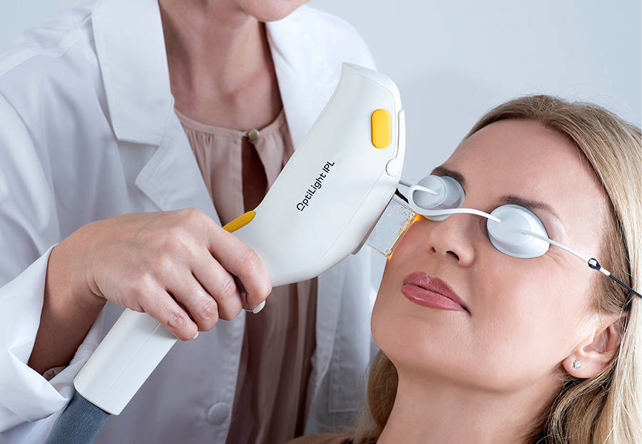
Millions of people suffer from eye and vision-related problems. For some, the solution is as simple as wearing prescription eyeglasses or contact lenses. However, others face challenges that aren’t as easy to rectify. That’s where Lumenis comes into play.
As a world leader in minimally invasive clinical solutions for both ophthalmology and aesthetic markets, it’s making a significant difference. Not only does Lumenis develop advanced energy-based technologies, but it also commercializes them.
The company continues to find innovative solutions. However, it’s already provided the fields of ophthalmology and aesthetic with remarkable technologies. OptiLight is just one example.
Eye and Vision-Related Solution
The company invented the first and only technology to treat individuals for dry eye disease caused by MGD. Not only is it approved by the Food and Drug Administration (FDA) but also patented. Optimal Pulse Technology (OPT) makes it easier to successfully manage this particular eye disease. Overall, OptiLight is safe, precise, comfortable, and effective.
Managing Symptoms
According to recent statistics, roughly 22% of the U.S. population suffers from dry eye disease along with MGD. That combination leads to an array of uncomfortable symptoms.
Dry eye occurs when a person’s eyes don’t produce adequate tears to keep them lubricated or when tears don’t work the way they should. MGD is a condition that affects the small glands in the eyelid responsible for making the oil layer for tears.
Having to deal with one of these problems is bad enough. However, living with both can prove debilitating for some people. Thanks to OptiLight, people with the two conditions get much-needed relief. Here are the ways this advanced technology helps:
Reduces inflammatory mediators, which, thereby, prevents inflammation
Improves tear breakup time that, in response, decreases osmolarity
Alleviates abnormal blood vessels that commonly cause inflammation
Restores meibomian glands so they function properly
Decreases demodex mites that cause infection and the accumulation of bacteria on the eyelids
What Does OptiLight Consist Of?
As part of this revolutionary system, ophthalmologists and optometrists can select different devices based on their needs.
Patented OPT Handpiece
Not two people have identical faces. Even among twins, facial contours differ, even if slightly. To ensure excellent results, this handpiece allows ophthalmologists and optometrists to customize treatments to cover every curve.
Ergonomic IPL Handpiece
To treat a broader area, Lumenis recommends this option. Patented with SapphireCool technology, it breaks the cycle of inflammation associated with dry eye disease caused by MGD.
Opti-Tip
For maximum energy control, the Opti-Tip focuses light energy on more delicate areas. As with the other Lumenis devices, it’s safe and effective. At the same time, the Opti-Tip is 100% hygienic.
Key Benefits of OptiLight
OptiLight utilizes embedded settings that adhere to strict protocols, making it safe and effective tool.
Additionally, they use Optimal Pulse Technology (OPT), which transforms light-based therapy. As a result, ophthalmologists and optometrists can treat the eyes with absolute precision and control. That’s because while this technology provides optimal energy, it never has spike inconsistencies.
Doctors can treat a patient in just 15 minutes. Specifically for dry eye disease with MGD, it provides an improvement in just four sessions.
The Bottom Line
Lumenis dedicated years to develop OptiLight. Now, eye doctors can incorporate a system into their practice to provide better patient care.

LASIK co-management is an integral part of vision correction surgery that often goes unnoticed. It involves a partnership between your primary eye care professional and your LASIK surgeon. This cooperative approach ensures that you're provided with the best possible care before, during, and after your procedure.
LASIK co-management is the collaborative effort of your eye care team to ensure your vision correction procedure is safe and successful. It's a team approach where everyone plays a significant role in your eye health journey.
The Evaluation Process
The evaluation process in LASIK co-management is thorough and meticulous. It begins with a comprehensive eye examination by your primary care optometrist. This examination is meant to ascertain your eye health status and determine if you're an appropriate candidate for the LASIK procedure.
The evaluation process underscores the collaborative nature of LASIK co-management. Every step is carefully coordinated to ensure your eye health is prioritized and that you're receiving the best possible care.
The Importance of Consultation in LASIK Co-management
Consultation is a crucial aspect of LASIK co-management. It allows for patient education, addressing concerns and questions, and establishing a clear understanding of the expected outcomes.
During the consultation phase, your optometrist will explain the results of your eye examination and how it relates to your suitability for LASIK surgery. They will also discuss the risks and benefits of the procedure, giving you a balanced perspective to make an informed decision.
The consultation with your LASIK surgeon will delve into the specifics of the operation, including the technology used, the surgical process, and the anticipated recovery timeline. Having the opportunity to consult with both your optometrist and your surgeon ensures that all your concerns are addressed, allowing you to proceed with confidence.
The Role of Communication
Effective communication is the backbone of successful LASIK co-management. It facilitates a seamless transition from your optometrist to your surgeon, creating a cohesive care plan that's centered around your specific needs.
This communication is not just between healthcare providers but also involves the patient. Regular updates on your progress, detailed explanations about each stage of the process, and addressing any concerns or questions you may have are all essential parts of the communication process.
Post-care in LASIK Co-management
Effective post-care in LASIK co-management is crucial in ensuring a successful outcome and speedy recovery. Post-care involves monitoring your healing progress, managing any discomfort or side effects, and ensuring that your vision is improving as expected.
Your optometrist plays a key role in this post-care. They will schedule follow-up visits to check your eyes and ensure they are healing correctly. They will also provide guidance on activities you should avoid and actions you can take to promote healing.
Additionally, if there are any complications or concerns, your optometrist is your first point of contact. They will liaise with your surgeon to address these concerns, demonstrating once again the importance of the collaborative approach in LASIK co-management.
The Benefits of LASIK Co-management
There are many benefits of LASIK co-management. First, it provides a comprehensive approach to your eye care, combining the expertise of your optometrist and your surgeon. This ensures that you're receiving the most thorough care possible.
Second, LASIK co-management creates a seamless patient experience. Your care transitions smoothly from your optometrist to your surgeon and back again. This eliminates any confusion or stress that can often come with navigating healthcare systems.
Finally, LASIK co-management provides continuity of care. Your optometrist, who is already familiar with your eye health history, remains involved in your care throughout the LASIK process. This continuity not only enhances your comfort level but also contributes to a better overall outcome.
Begin Your Journey to Clear Vision with LASIK Co-management Today
LASIK co-management represents the gold standard in vision correction procedures. It leverages the combined expertise of your optometrist and your surgeon, creating a streamlined, cohesive, and patient-centered approach to your care.
From your initial evaluation to post-operative care, every step of the process is carefully coordinated to ensure your safety, comfort, and the success of your procedure. And with the myriad benefits of LASIK co-management, you can approach your vision correction journey with confidence and peace of mind.
If you're considering LASIK surgery, choose a provider that offers LASIK co-management. It's a proven approach that puts your needs at the center and prioritizes your eye health at every step.

A refraction test, also called a vision test, is usually performed as a part of a routine eye examination. The purpose of this test is to determine if a person has a refractive error which would then mean the patient would need glasses or contact lenses.
What Is The Normal Value for Refraction Test?
A value of 20/20 is normal (optimum) vision. This means that individuals who have 20/20 vision are able to read letters that are 3/8-inch (1 centimeter) tall from 20 feet (6 meters) away. The normal uncorrected vision (without glasses or contact lenses) refractive error is zero (plano). Individuals who don’t have 20/20 vision, have what is called a refractive error. A refractive error means that the light is not bending properly when it passes through the lens of the eye. The refraction test will tell the doctor what prescription lens should be used in order to have 20/20 vision.
For people over age 40 who have normal distance vision but difficulty with near vision, a refraction test with a small type size is used to determine normal near vision and the correct power of reading glasses.
How Is The Refraction Test Performed?
The test is performed by having the patient seated in a chair that has a special device (called a phoropter or refractor) attached to it. The patient looks through the device and focuses on an eye chart 20 feet (6 meters) away. The device contains lenses of different strengths that can be moved into the patient’s view. The test is performed one eye at a time. If the patient is wearing contact lenses, they should be removed before the test.
In case the final vision is less than 20/20 even with lenses, then there is probably another non-optical problem with the eye. The vision level achieved during the refraction test is called the best-corrected visual acuity (BCVA).
What Are The Causes of Abnormal Refraction Test Results?
Abnormal results may be due to:
Astigmatism (abnormally curved cornea causing blurred vision)
Hyperopia (farsightedness)
Myopia (nearsightedness)
Presbyopia (inability to focus on near objects that develop with age)
Other conditions under which the test may be performed:
Corneal ulcers and infections
Loss of sharp vision due to macular degeneration
Retinal detachment (separation of the light-sensitive membrane (retina) in the back of the eye from its supporting layers)
Retinal vessel occlusion (blockage in a small artery that carries blood to the retina)
Retinitis pigmentosa (an inherited disorder of the retina)
There is an art to refraction and the optometrist will always answer the patient’s questions and as well as discuss their findings. Based on the results of the refraction test, they can determine the amount of myopia, hyperopia or astigmatism.

The parts of a comprehensive eye examination vary according to the patient's age, date of last exam, and other factors. Not all parts of the eye exam may be needed or performed, but the first part of the eye exam will include documenting medical history. Here are some eye and vision tests that are likely to be encountered during a comprehensive eye exam:
Visual Acuity Tests
Visual acuity tests measure the sharpness of vision and are usually performed using a projected eye chart to measure the distance visual acuity and a hand-held small acuity chart to measure the near vision (for reading).
Color Blindness Test
A screening test that checks the color vision is often performed early in a comprehensive eye exam to rule out color blindness.
Cover test to check eye alignment.
A test used to assess strabismus or a more subtle binocular vision problem that could cause eye strain or amblyopia (lazy eye).
Ocular Motility (Eye Movements) Testing
Ocular motility testing is performed to determine how well eyes can follow a moving object and/or quickly move between and accurately fixate on two separate targets.
Stereopsis (Depth Perception) Test
This is used to test perception of depth and 3-dimensional structure obtained on the basis of visual information deriving from two eyes by individuals with normally developed binocular vision.
Retinoscopy
This test is used to estimate which lens powers will best correct distance vision. Based on the way the light reflects from the eye, the doctor is able to obtain an approximation of the eyeglass prescription. This test is useful for children and patients who are unable to accurately answer the doctor's questions.
Manual refraction with a phoropter.
This is the test used to determine the exact eyeglass prescription.


Interested in our
Services?
Request appointment with this form to schedule a time with our professional staff!
- Monday8:00 AM - 7:00 PM
- Tuesday8:00 AM - 5:00 PM
- Wednesday8:00 AM - 5:00 PM
- Thursday8:00 AM - 7:00 PM
- Friday8:00 AM - 5:00 PM
- Saturday8:00 AM - 4:00 PM
- SundayClosed
Powered by: 
Unfortunately, the global supply chain problems are affecting contact lens orders. Manufacturers are reporting delays from two weeks to three months. We still have select contact lens supplies in stock and will do whatever possible to accommodate your needs. We also recommend reordering your contact lenses three months in advance of the use of your last pair.
ORDER CONTACT LENSES